An ultramicrotome that transforms a normal SEM to a volume SEM
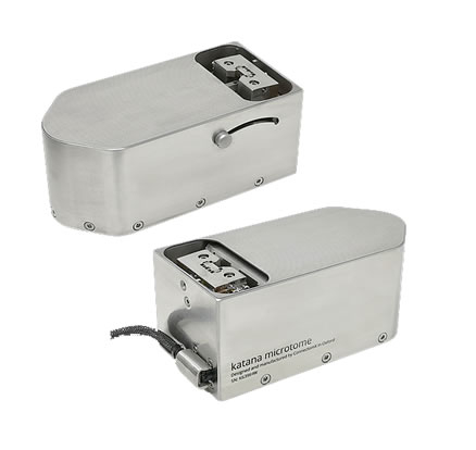
At a mere 56 mm in height, katana ultramicrotome is designed to fit inside the vacuum chamber of many SEMs. Easy installation has been at the heart of our design. The microtome can be attached to/remove from an SEM stage by a lever mechanism. All electronic signals are fed through a vacuum flange via a single connector. The user friendly design enables a quick and easy switch from your normal SEM to a volume SEM and back.
Serial block-face SEM
In serial block-face imaging, a microtome resides inside the vacuum chamber of an SEM. A diamond knife repeatedly removes a thin surface layer from the sample block. The removed layer can be as thin as 15 nm. After each sectioning, the exposed block surface is imaged. This automated in situ method can acquire a series of electron micrographs over a large volume.
Ultra is also for ultrasonic
Think about the sawing motion you make when slicing a loaf of bread, the sample cutting force can be greatly reduced - and the surface texture of the cut improved - by oscillating the diamond knife at ultrasonic speeds. The FEA optimised knife design enables a piezo driven in-plane oscillation at an amplitude up to 170 nm at resonant frequency. The oscillation amplitude is constantly measured by a sensing piezo. The dual-piezo design allows the fine tuning of the oscillation mode which varies between different knife holders and samples.
Without knife oscillation, softer pigment granules are pulled out of the block by the knife. This behaviour is immediately prevented when oscillation is turned on.
Nanometre precision
A precise and repeatable stepping motion in Z-direction is crucial to produce accurate Z scaling in final 3D volume data. The position of our sample stage - driven by an ultra-fast motor - is measured constantly by a nanometre resolution encoder so that any drift can be instantly corrected.
The accompanied benefits are:
- Debris mitigation: as the knife retracts after each cut, the sample can drop hundreds of microns away from the removed material. This reduces the chance of debris falling onto the block surface by electrostatic force;
- Reduced working distance: samples can move closer to the detector during imaging, thereby improving the collection of backscattered electrons and thus signal-to-noise ratio.
Finding cutting plane made easy
For someone who is new to ultramicrotomy, finding the cutting plane is often a daunting experience. Our carefully designed digital viewer makes this process simple and smooth. The digital viewer sits over the microtome so that the approach process can be done on an SEM stage. It has a 40 fps 2.3 MP CMOS camera coupled with a high resolution lens to create a crisp and low latency view of the knife and sample. A series of internal mirrors tracks the movement of the knife so that the blade of the diamond always stays focused and stationary on the screen.
Open ad friendly to users
Katana microtome is controlled via a tablet with a familiar multi-touch user interface that enables you to control the microtome with your fingertip. It communicates with our universal SEM automation software which works with existing SEM control software to conduct scanning, imaging monitoring and image saving automatically. Soon, katana microtome will be fully integrated with SBEMimage, a versatile acquisition control software for serial block-face electron microscopy developed by The Friedrich Miescher Institute for Biomedical Research.
We have always loved using open source and we believe in giving back to open source as much as we benefit from it. You can control the microtome via USB by sending serial commands which are available to users. You can build your own apps to control the microtome, whether it be a from simple cutting and actuating process, to complex automation with other instruments.